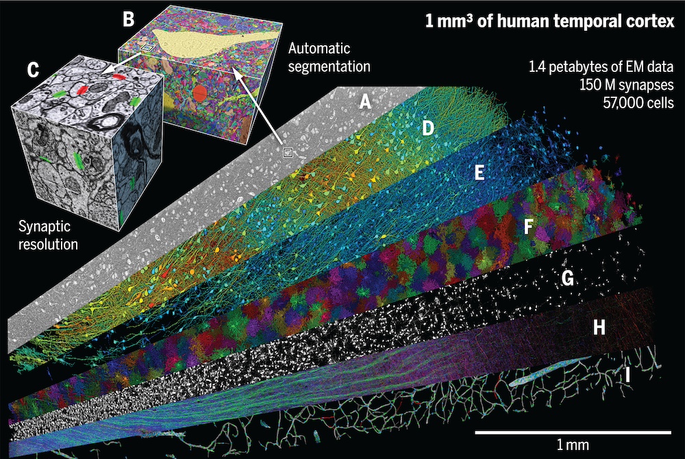
A petavoxel fragment of human cerebral cortex reconstructed at nanoscale resolution
Science, 2024.
A complete understanding of the human brain begins with elucidation of its structural properties at a subcellular level. To provide a valuable resource for the scientific community and to better understand the structure of the human temporal cortex, Shapson-Coe et al. performed an electron microscopy reconstruction of a cubic millimeter of human temporal cortex. The authors produced 1.4 petabytes of electron microscopy data; classified and quantified cell types, vessels and synapses; and developed a freely available tool for analyzing these data. Their findings allowed the authors to identify previously unknown aspects of the human temporal cortex.
Acknowledgements
We thank M. Frosch and S. Huang for help with obtaining andcharacterizing the sample of brain tissue used in this study; the multi SEM team at Carl Zeiss Microscopy who helped us get their device into our workflow; K. Rockland for helping us with relevant literature; J. Choi for help with the CAVE instantiation; students A. Judd, E. Pavarino, R. Jiang, R. Han, P. Miao, T. Lu, and J. Afeeli for help with various aspects of this project; and S. Montgomery for careful reading of the manuscript. Funding: This work was supported by the National Institute of Mental Health of the National Institutes of Health (Conte Center award P50MH094271 to J.W.L.); the Stanley Center at the Broad Institute (J.W.L.); the BRAIN Initiative of the National Institutes of Health (grants UG3MH123386, U19 NS104653, U24 NS109102, and UO1 EB026996 to J.W.L.); the National Science Foundation (grant NCS-FO-2124179 to H.P. and J.T. and NeuroNex 2 award 2014862 to F.C.); the National Institutes of Health (grant RF1MH125932 to F.C.); and Intelligence Advanced Research Projects Activity through the Department of Interior/Interior Business Center (contract D16PC00004 and D16PC0005 to F.C.).