Frenet-Serret Frame-based Decomposition for Part Segmentation of 3D Curvilinear Structures
IEEE Transactions on Medical Imaging, 2025.
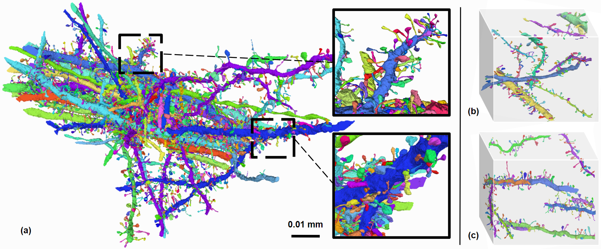
Accurate segmentation of anatomical substructures within 3D curvilinear structures in medical imaging remains challenging due to their complex geometry and the scarcity of diverse, large-scale datasets for algorithm development and evaluation. In this paper, we use dendritic spine segmentation as a case study and address these challenges by introducing a novel Frenet–Serret Frame–based Decomposition, which decomposes 3D curvilinear structures into a globally smooth continuous curve that captures the overall shape, and a cylindrical primitive that encodes local geometric properties. This approach leverages Frenet–Serret Frames and arc length parameterization to preserve essential geometric features while reducing representational complexity, facilitating data-efficient learning, improved segmentation accuracy, and generalization on 3D curvilinear structures. To rigorously evaluate our method, we introduce two datasets: CurviSeg, a synthetic dataset for 3D curvilinear structure segmentation that validates our method’s key properties, and DenSpineEM, a benchmark for dendritic spine segmentation, which comprises 4,476 manually annotated spines from 70 dendrites across three public electron microscopy datasets, covering multiple brain regions and species. Our experiments on DenSpineEM demonstrate exceptional cross-region and cross-species generalization: models trained on the mouse somatosensory cortex subset achieve 94.43% Dice, maintaining strong performance in zero-shot segmentation on both mouse visual cortex (95.61% Dice) and human frontal lobe (86.63% Dice) subsets. Moreover, we test the generalizability of our method on the IntrA dataset, where it achieves 77.08% Dice (5.29% higher than prior arts) on intracranial aneurysm segmentation from entire artery models. These findings demonstrate the potential of our approach for accurately analyzing complex curvilinear structures across diverse medical imaging fields.
Acknowledgements
This work was partially supported by NSF grant NCS-FO-2124179, and NIH grant 1U01NS132158. Leslie Gu is also supported by SEAS Graduate Fellowship. Donglai Wei is supported by NSF-IIS 2239688.