Vials: Visualizing Alternative Splicing of Genes
IEEE Transactions on Visualization and Computer Graphics, 2016.
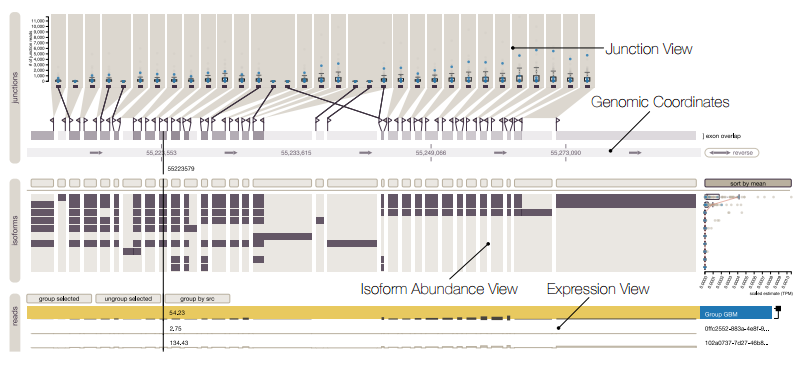
Alternative splicing is a process by which the same DNA sequence is used to assemble different proteins, called protein isoforms. Alternative splicing works by selectively omitting some of the coding regions (exons) typically associated with a gene. Detection of alternative splicing is difficult and uses a combination of advanced data acquisition methods and statistical inference. Knowledge about the abundance of isoforms is important for understanding both normal processes and diseases and to eventually improve treatment through targeted therapies. The data, however, is complex and current visualizations for isoforms are neither perceptually efficient nor scalable. To remedy this, we developed Vials, a novel visual analysis tool that enables analysts to explore the various datasets that scientists use to make judgments about isoforms: the abundance of reads associated with the coding regions of the gene, evidence for junctions, i.e., edges connecting the coding regions, and predictions of isoform frequencies. Vials is scalable as it allows for the simultaneous analysis of many samples in multiple groups. Our tool thus enables experts to (a) identify patterns of isoform abundance in groups of samples and (b) evaluate the quality of the data. We demonstrate the value of our tool in case studies using publicly available datasets.