Imaging a 1 mm3 Volume of Rat Cortex Using a MultiBeam SEM
Cambridge Univ Press: Microscopy and Microanalysis, 2016.
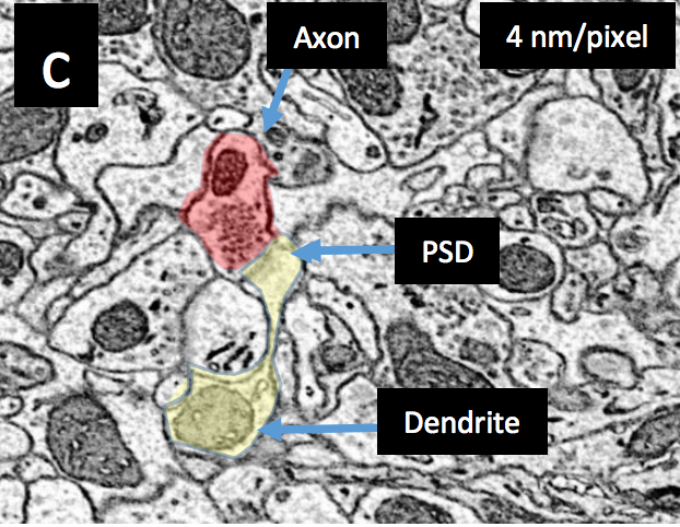
The rodent brain is organized with length scales spanning centimeters to nanometers —6 orders of magnitude [1]. At the centimeter scale, the brain consist of lobes of cortex, the cerebellum, the brainstem and the spinal cord. The millimeter scale have neurons arranged in columns, layers, or otherwise clustered. Recent technological imaging advances allow the generation of neuronal datasets spanning the spatial range from nanometers to 100s of microns [2,3]. Collecting a 1 mm3 volume dataset of brain tissue at 4 nm x-y resolution using the fastest signal-beam SEM would require ~6 years. To move to the next length and volume scale of neuronal circuits requires several technological advances. The multibeam scanning electron microscope (mSEM) represents a transformative imaging technology that enables neuroscientists to tackle millimeter scale cortical circuit problems. In this work we describe a workflow from tissue harvest to imaging that will generate a 2 petabyte dataset (> 300,000,000 images) of rat visual cortex imaged at a 4nm x 4nm x-y (Nyquist sampling of membranes) and 30nm section thickness in less than 6 months.